Anatomy Rib Cage / Rib Cage Drawing Hd Stock Images Shutterstock : Shaped somewhat like a cone, it is created by the individual ribs connecting to the spine above and to the sternum below.
byAdmin•
0
Anatomy Rib Cage / Rib Cage Drawing Hd Stock Images Shutterstock : Shaped somewhat like a cone, it is created by the individual ribs connecting to the spine above and to the sternum below.. The upper edge is round and the lower sharp. #proko #art #anatomy #ribs #ribcage #humananatomy #tutorial. Diagram of human body, liver rib cage, rib cage diagram labeled, rib cage diagram numbered, rib cage diaphragm, rib cage heart, rib cage organs anatomy, rib cage pain, stomach, diagram of human body, liver rib cage, rib cage diagram labeled, rib cage diagram numbered, rib cage diaphragm, rib cage. The thoracic cage (rib cage) is the skeletal framework of the thoracic wall, which encloses the thoracic cavity. At the chest, many rib bones connect to the sternum via costal cartilage,.
The primary causes of pain under the left rib cage. In this image, you will find clavicle, true ribs, sternal angle, costal cartilage, false ribs, floating ribs, seventh cervical vertebra, first thoracic vertebra, jugular notch, manubrium, body, xiphoid process, sternum in it. Human muscles · april 17, 2020. The spleen is used to filter red blood cells and hangs in the upper part of the abdomen. Click the image to watch the anatomy of the rib cage video.
Rib Cage 3d Models For Download Turbosquid from static.turbosquid.com Diagram of human body, liver rib cage, rib cage diagram labeled, rib cage diagram numbered, rib cage diaphragm, rib cage heart, rib cage organs anatomy, rib cage pain, stomach, diagram of human body, liver rib cage, rib cage diagram labeled, rib cage diagram numbered, rib cage diaphragm, rib cage. On the interior wall of the rib body is a channel, sulcus costae, with blood vessels and nerves. The rib cage is a bony structure found in the chest (thoracic cavity). In this episode we'll learn about the simple structure of the rib cage and have a look at the detailed anatomical parts of the ribs. Each pair is numbered based on their attachment to the sternum, a bony process at the front of the rib cage which serves as an anchor point. The cartilage strips are called costal cartilage (costal is the anatomical adjective that refers to the rib) and connect on their other end to the sternum. Both are given some protection by the rib bones. An enlarged or ruptured spleen can cause sudden or chronic pain under the left rib cage that ends up migrating towards the back and/or shoulders.
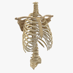
The cartilage strips are called costal cartilage (costal is the anatomical adjective that refers to the rib) and connect on their other end to the sternum. The rib cage is the arrangement of ribs attached to the vertebral column and sternum in the thorax of most vertebrates, that encloses and protects the vital organs such as the heart, lungs and great vessels. The rib cage, shaped in a mild cone shape and more flexible than most bone sets, is made up of varying elements such as the thoracic vertebra, 12 equally paired ribs, costal cartilage, and held together anteriorly by the sternum. At the chest, many rib bones connect to the sternum via costal cartilage,. A rib has a flat body, as you can see from the picture of the anatomy of the human rib cage. The thoracic back describes the area of your back from just below the base of your skull to about 5 inches below the lower part of your shoulder blades. They are extremely light, but highly resilient; The rib cage is a bony structure found in the chest (thoracic cavity). It is made up of 12 pairs of ribs. Anatomy of the rib cage. Both are given some protection by the rib bones. Diagram of human body, liver rib cage, rib cage diagram labeled, rib cage diagram numbered, rib cage diaphragm, rib cage heart, rib cage organs anatomy, rib cage pain, stomach, diagram of human body, liver rib cage, rib cage diagram labeled, rib cage diagram numbered, rib cage diaphragm, rib cage. Animated full human body anatomy.
Contributing to their role in protecting the internal thoracic organs. Check out our anatomy rib cage selection for the very best in unique or custom, handmade pieces from our shops. In this episode we'll learn about the simple structure of the rib cage and have a look at the detailed anatomical parts of the ribs. Rib cage pain can be caused. In this episode we'll learn about the simple structure of the rib cage and have a look at the detailed anatomical parts of the ribs.
Rib Cage Human Skeleton Sternum Anatomy Adan Anatomy Human Body Png Pngegg from e7.pngegg.com Clavicle anatomy and rib cage anatomy. The cartilage strips are called costal cartilage (costal is the anatomical adjective that refers to the rib) and connect on their other end to the sternum. The ribs are a set of twelve paired bones which form the protective 'cage' of the thorax. In this image, you will find clavicle, true ribs, sternal angle, costal cartilage, false ribs, floating ribs, seventh cervical vertebra, first thoracic vertebra, jugular notch, manubrium, body, xiphoid process, sternum in it. Ten of the twelve ribs connect to strips of hyaline cartilage on the anterior side of the body. The thoracic cage consists of the 12 thoracic vertebrae, the associated intervertebral discs, 12 pairs of ribs with their costal cartilages, and the sternum. An enlarged or ruptured spleen can cause sudden or chronic pain under the left rib cage that ends up migrating towards the back and/or shoulders. The thoracic cage (rib cage) is the skeletal framework of the thoracic wall, which encloses the thoracic cavity.
The thoracic back describes the area of your back from just below the base of your skull to about 5 inches below the lower part of your shoulder blades.
Cavea thoracis, thoracic cage, rib cage, rib cage, thoracic cage, thoracic cage structure (body structure), thoracic cage structure, rib cage, nos. In this image, you will find clavicle, true ribs, sternal angle, costal cartilage, false ribs, floating ribs, seventh cervical vertebra, first thoracic vertebra, jugular notch, manubrium, body, xiphoid process, sternum in it. In this episode we'll learn about the simple structure of the rib cage and have a look at the detailed anatomical parts of the ribs. The upper edge is round and the lower sharp. The top edge of the manubrium has a depression called the suprasternal or jugular notch. Ten of the twelve ribs connect to strips of hyaline cartilage on the anterior side of the body. The spleen is used to filter red blood cells and hangs in the upper part of the abdomen. Shaped somewhat like a cone, it is created by the individual ribs connecting to the spine above and to the sternum below. Diagram of human body, liver rib cage, rib cage diagram labeled, rib cage diagram numbered, rib cage diaphragm, rib cage heart, rib cage organs anatomy, rib cage pain, stomach, diagram of human body, liver rib cage, rib cage diagram labeled, rib cage diagram numbered, rib cage diaphragm, rib cage. The rib cage is a bony structure found in the chest (thoracic cavity). 16 photos of the rib cage diagram with organs. The thoracic cage (rib cage) is the skeletal framework of the thoracic wall, which encloses the thoracic cavity. A rib has a flat body, as you can see from the picture of the anatomy of the human rib cage.
The thoracic cage consists of the 12 thoracic vertebrae, the associated intervertebral discs, 12 pairs of ribs with their costal cartilages, and the sternum. It consists of the 12 pairs of ribs with their costal cartilages and the sternum (figure 6.38). Contributing to their role in protecting the internal thoracic organs. Each pair is numbered based on their attachment to the sternum, a bony process at the front of the rib cage which serves as an anchor point. In this image, you will find clavicle, true ribs, sternal angle, costal cartilage, false ribs, floating ribs, seventh cervical vertebra, first thoracic vertebra, jugular notch, manubrium, body, xiphoid process, sternum in it.
Human Rib Cage Anatomy Royalty Free Vector Image from cdn5.vectorstock.com In this episode we'll learn about the simple structure of the rib cage and have a look at the detailed anatomical parts of the ribs. They articulate with the vertebral column posteriorly, and terminate anteriorly as cartilage (known as costal cartilage). It consists of the 12 pairs of ribs with their costal cartilages and the sternum (figure 6.38). An enlarged or ruptured spleen can cause sudden or chronic pain under the left rib cage that ends up migrating towards the back and/or shoulders. Anatomy of the rib cage. #proko #art #anatomy #ribs #ribcage #humananatomy #tutorial. Cavidad costal, estructura de la caja torácica (estructura corporal), estructura de la caja torácica, jaula costal. The rib below that is rib 2, and it connects to the t2 thoracic vertebra, and so on.
The upper edge is round and the lower sharp.
Derived from the nih umls ( unified medical language. The sternum is a flat bone that is made up of three parts, the (1) manubrium, (2) body, and the (3) xiphoid process. Shaped somewhat like a cone, it is created by the individual ribs connecting to the spine above and to the sternum below. The ribs are a set of twelve paired bones which form the protective 'cage' of the thorax. The lungs are two separate but connected organs located in the upper chest, covered by the rib cage. In this image, you will find clavicle, true ribs, sternal angle, costal cartilage, false ribs, floating ribs, seventh cervical vertebra, first thoracic vertebra, jugular notch, manubrium, body, xiphoid process, sternum in it. Clavicle anatomy and rib cage anatomy. Rib cage, in vertebrate anatomy, basketlike skeletal structure that forms the chest, or thorax, and is made up of the ribs and their corresponding attachments to the sternum (breastbone) and the vertebral column. Click the image to watch the anatomy of the rib cage video. The thoracic cage (rib cage) forms the thorax (chest) portion of the body. A rib has a flat body, as you can see from the picture of the anatomy of the human rib cage. The spleen is used to filter red blood cells and hangs in the upper part of the abdomen. Ten of the twelve ribs connect to strips of hyaline cartilage on the anterior side of the body.